Pelvic Anatomy Ligaments - 1 1 Female Pelvis Vessels Muscles Nerves With Vessels Ligaments Muscles Nerves Removable Organs Medical Anatomical Model Biology Aliexpress - The inlet to the pelvic canal is at the level of the sacral promontory and superior aspect of the pubic bones.
Pelvic Anatomy Ligaments - 1 1 Female Pelvis Vessels Muscles Nerves With Vessels Ligaments Muscles Nerves Removable Organs Medical Anatomical Model Biology Aliexpress - The inlet to the pelvic canal is at the level of the sacral promontory and superior aspect of the pubic bones.. Imaios and selected third parties, use cookies or similar technologies, in particular for audience measurement. These ligaments are important stabilizers. The named ligaments of the pelvis mostly arise from the sacrum and attach to varying segments of the pelvic bone. • posterolateral wall—piriformis and coccygeus muscles. Additional ligaments may be found in the female pelvis.
There are two major groups of ligaments that provide nearly all the structure of the pelvis. It extends from the lateral pelvic walls on both sides, and folds over the internal female genitalia, covering their surface anteriorly and posteriorly. Bones and ligaments of the female pelvis. Pain caused by stretching the sacrospinous and sacroiliac ligaments is felt within the s1 dermatome from a point posterolateral to the hip. The outlet is formed by the pubic arch, ischial spines, sacrotuberous ligaments, and the coccyx.
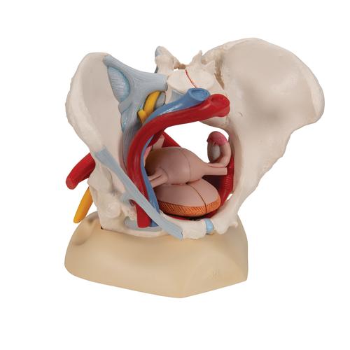
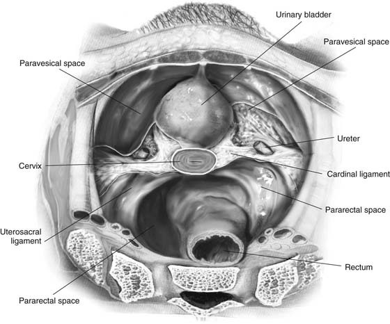
Anatomical Teaching Models Plastic Human Pelvic Models Female Pelvis With Ligaments Vessels Nerves Pelvic Floor Muscles And Organs from www.3bscientific.com Pain caused by stretching the iliolumbar ligament is referred to the inguinal region (the differential diagnosis includes a hip disorder). • anterolateral wall—hip bone and obturator internus muscles. Additional ligaments may be found in the female pelvis. Pelvic anatomy sacrouterine ligament cardinal ligaments pelvic fascia sacrospinous ligament urethral support bladder support rectal support. The three bony structures (ilium, ischium, and pubis) are held together with strong ligaments that are important in understanding pelvic anatomy and biomechanics. The cardinal ligaments, also known as the transverse cervical ligaments, the lateral cervical ligaments, or mackenrodt's ligaments, are fibrous bands that attached the cervix to the lateral pelvic walls. This is part of the forced closure method that the pelvis adopts in order to keep itself secure. The broad ligament is a flat sheet of peritoneum, associated with the uterus, fallopian tubes and ovaries.
The pectineal ligament is usually around 6 cm long in adults.
Pelvic ligaments tests is considered positive in these situations: Functional anatomy of the male pelvic floor online course: The pelvis itself is a paired composite structure made up by three bones (ilium, ischium and pubis) that articulates with the sacral part of the axial spine. The outlet is formed by the pubic arch, ischial spines, sacrotuberous ligaments, and the coccyx. Pelvic anatomy sacrouterine ligament cardinal ligaments pelvic fascia sacrospinous ligament urethral support bladder support rectal support. Other ligaments attached to bony pelvis include the sacrococcygeal ligaments, pubic symphysis ligaments, and endopelvic fascia ligament. The pectineal ligament is usually around 6 cm long in adults. This is part of the forced closure method that the pelvis adopts in order to keep itself secure. The femoral ligaments act to stabilize the ball and socket joint of the hip, connecting to the ilium and the ischium. Pain caused by stretching the iliolumbar ligament is referred to the inguinal region (the differential diagnosis includes a hip disorder). The vertebropelvic ligaments include the iliolumbar, sacrotuberous and sacrospinous ligaments. The broad ligament can be further divided into three components. Two innominate bones, which consist of the:
The broad ligament can be further divided into three components. Lets get deeper into the musculoskeletal anatomy of the hip and look at the bones and bony bits of the pelvis, and the ligaments that attach here and hold it. The 3 groups of ligaments are: Pain caused by stretching the sacrospinous and sacroiliac ligaments is felt within the s1 dermatome from a point posterolateral to the hip. The ilium, ischium and the pubic bone.
Intra Abdominal Pelvic Anatomy Obgyn Key from obgynkey.com Pelvic ligaments tests is considered positive in these situations: The broad ligament is a flat sheet of peritoneum, associated with the uterus, fallopian tubes and ovaries. It extends to both sides of the pelvic wall. Pelvic anatomy sacrouterine ligament cardinal ligaments pelvic fascia sacrospinous ligament urethral support bladder support rectal support. The ligaments of the pelvis, are amongst the strongest in the human body. The outlet is formed by the pubic arch, ischial spines, sacrotuberous ligaments, and the coccyx. It extends from the lateral pelvic walls on both sides, and folds over the internal female genitalia, covering their surface anteriorly and posteriorly. These are accessory ligaments to the sacroiliac joints of the pelvis and are important in maintaining its stability.
They form what can be described as a basket weave formation, in order to create strength and tensegrity within the structure.
The inlet to the pelvic canal is at the level of the sacral promontory and superior aspect of the pubic bones. Additional ligaments may be found in the female pelvis. The pelvis itself is a paired composite structure made up by three bones (ilium, ischium and pubis) that articulates with the sacral part of the axial spine. Other ligaments attached to bony pelvis include the sacrococcygeal ligaments, pubic symphysis ligaments, and endopelvic fascia ligament. Resist shear and flexion forces. Female pelvis ppt by mayil rasamani), which are reflections of the broad ligament attaching the ovaries to the lateral pelvis. There are many organs that sit in the pelvis, including much of the urinary system, and lots of the male or female reproductive systems. The ligaments of the pelvis, are amongst the strongest in the human body. This will be explored further on. • posterolateral wall—piriformis and coccygeus muscles. Bones and ligaments of the female pelvis. The inlet to the pelvic canal is at the level of the sacral promontory and superior aspect of the pubic bones.; The pelvis is a boney structure at the base of the lumbar spine.
Cookies allow us to analyze and store information such as the characteristics of your device as well as certain personal data (e.g., ip addresses, navigation, usage or geolocation data, unique identifiers). Additional ligaments may be found in the female pelvis. The three bony structures (ilium, ischium, and pubis) are held together with strong ligaments that are important in understanding pelvic anatomy and biomechanics. The femoral ligaments act to stabilize the ball and socket joint of the hip, connecting to the ilium and the ischium. The pelvis itself is a paired composite structure made up by three bones (ilium, ischium and pubis) that articulates with the sacral part of the axial spine.
References In Surgical Exposure And Anatomy Of The Female Pelvis Surgical Clinics from els-jbs-prod-cdn.jbs.elsevierhealth.com It is close to the major vasculature of the pelvis, including external iliac vein. The vertebropelvic ligaments include the iliolumbar, sacrotuberous and sacrospinous ligaments. It extends from the lateral pelvic walls on both sides, and folds over the internal female genitalia, covering their surface anteriorly and posteriorly. Pain caused by stretching the iliolumbar ligament is referred to the inguinal region (the differential diagnosis includes a hip disorder). The broad ligament is a flat sheet of peritoneum, associated with the uterus, fallopian tubes and ovaries. • anterolateral wall—hip bone and obturator internus muscles. Pelvic floor consists of two ligaments: These ligaments are important stabilizers.
The pelvis is a boney structure at the base of the lumbar spine.
The outlet is formed by the pubic arch, ischial spines, sacrotuberous ligaments, and the coccyx. The enclosed space between the inlet and outlet is called the true pelvis, with. The pelvis is a boney structure at the base of the lumbar spine. • anterolateral wall—hip bone and obturator internus muscles. The outlet is formed by the pubic arch, ischial spines, sacrotuberous ligaments, and the coccyx. Below the pelvic brim), posterior. Additional ligaments may be found in the female pelvis. Additional ligaments may be found in the female pelvis. Pain caused by stretching the iliolumbar ligament is referred to the inguinal region (the differential diagnosis includes a hip disorder). Female pelvis ppt by mayil rasamani), which are reflections of the broad ligament attaching the ovaries to the lateral pelvis. It extends from the lateral pelvic walls on both sides, and folds over the internal female genitalia, covering their surface anteriorly and posteriorly. • also known as pelvic cavity. The pelvic girdle and pelvic spine.
There are many organs that sit in the pelvis, including much of the urinary system, and lots of the male or female reproductive systems pelvic anatomy. • located inferior to the pelvic brim.


0 Komentar